Lung nodules are classified as either non-cancerous or cancerous. The most prevalent causes of benign nodules are granulomas (clumps of inflammatory tissue) and hamartomas (abnormal growths of connective tissue). The most prevalent causes of diseased or malignant lung nodules are lung cancer or cancer that has progressed from another part of the body to the lungs. Commonly, lung cancer is caused by cancerous or cancerous lung nodules.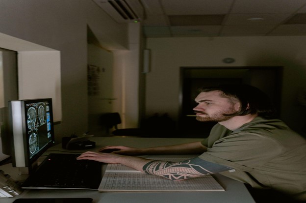
Generally speaking, pulmonary nodules may be categorized into a few broad categories:
- Tumors that are not cancerous, such as hamartomas, infections, such as bacterial diseases such as TB and fungal infections
- Malignant tumors, such as lung cancer and cancer, have metastasized to the lungs after spreading to other organs in the body.
Overall, the chance of a lung cluster being malignant is roughly 40%, although the likelihood of a lesion being cancerous varies significantly based on a variety of factors, such as:
- People under the age of 35 developing lung nodules are quite rare. Approximately half of all lung nodules in persons over the age of 50 are cancerous.
- Lung lesions that are calcified (https://www.healthline.com/health/calcified-granuloma) have a higher chance of being non-cancerous.
- It is more probable that nodules that are defined as “cavitary,” which means that the internal area of the nodule seems to retain air on X-rays, are non-cancerous.
- Compared to benign lung nodules, cancerous lung nodules tend to develop more quickly, with an average doubling period of roughly four months, while benign growths tend to stay relatively stable in size over time.
- Having a family history of cancer raises the likelihood that the tumor will be malignant.
- Some occupational exposures increase the probability that a nodule may turn out to be cancerous in the future.
- Smokers, both current and past, have a higher risk of developing malignant lung nodules than nonsmokers.
- Nodules that are larger in size seem to be more probable to be malignant than tiny nodules.
Common Causes Of Lung Nodules
Nodules that are smooth and spherical are more probable to be benign, but nodules that are uneven or “spiculated” are more prone to be malignant.
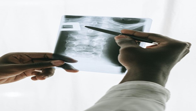
A prominent cause of lung nodules, tumors, and cavities continues to be infectious diseases. To establish a prognosis, discriminate between malignancy and pathogenic causes, as well as provide microbiological evidence for characterization and analysis, it is necessary to collect tissue samples in a safe environment. Tissue samples might be collected using bronchoscopy, percutaneous biopsy, or surgical biopsy, among other methods. Breath examination is the most risk-free method of preventing the sequelae of pleural and chest wall puncture.
The surgical robot that has been built has excellent control precision and can meet all of the criteria for usage. The well-established lesion location approach may give a high degree of accuracy for robot-assisted puncture surgery, which is advantageous. The omnidirectional needle placement technique that has been created may be used to find the most appropriate insertion site and insertion posture. A theoretical basis for further research in route planning or autonomous placement may be derived from this work, which might be useful for future research.
Tomographic Imaging Of Lung Nodules
When it comes to a specific diagnosis involving lung nodules, tomographic imaging is required. As you are undoubtedly aware, this form of imaging allows you, as a medical practitioner, to view inside the person’s body to determine whether or not the malignancy is there and whether or not it has spread.
A CAT scan would be the first thing that any specialist will request in order to examine the interior of the patient’s body and determine whether or not the cancer is present and has spread. So, this is among the most fundamental tests that you definitely must have while working in this field and in the detection of suspected lung cancer. Then there is the PET scan, and it can be quite beneficial in the process of detection.
Tool In Lesion Confirmation In Biopsies
The CABT Tool, or confirming biopsy tool, is fundamental to tools-in-lesions confirmation.
A pre-op CT is prepped and loaded. The lesion and the boundaries of the lesion are marked, and the system quickly learns the structure of the lung, including identifying possible paths to the lesion on the lung.
The Traditional Bronchoscopy Method
The diagnostic yield of traditional bronchoscopy, on the other hand, is low in the case of tiny, peripheral lesions. For more information on bronchoscopy, please click here. In electromagnetic navigation bronchoscopy (ENB), the bronchoscope and working channel are directed along the bronchial tree in order to reach a peripheral lesion with pinpoint accuracy.
In peripheral lesions, particularly tiny lesions, it has a significant impact on the diagnostic yield, and its significance has expanded beyond diagnosis to include therapeutic enablement as well as direct therapy.


